Synthesis of vancomycin fluorescent probes that retain antimicrobial ... - Nature.com
Abstract
Antimicrobial resistance is an urgent threat to human health, and new antibacterial drugs are desperately needed, as are research tools to aid in their discovery and development. Vancomycin is a glycopeptide antibiotic that is widely used for the treatment of Gram-positive infections, such as life-threatening systemic diseases caused by methicillin-resistant Staphylococcus aureus (MRSA). Here we demonstrate that modification of vancomycin by introduction of an azide substituent provides a versatile intermediate that can undergo copper-catalysed azide−alkyne cycloaddition (CuAAC) reaction with various alkynes to readily prepare vancomycin fluorescent probes. We describe the facile synthesis of three probes that retain similar antibacterial profiles to the parent vancomycin antibiotic. We demonstrate the versatility of these probes for the detection and visualisation of Gram-positive bacteria by a range of methods, including plate reader quantification, flow cytometry analysis, high-resolution microscopy imaging, and single cell microfluidics analysis. In parallel, we demonstrate their utility in measuring outer-membrane permeabilisation of Gram-negative bacteria. The probes are useful tools that may facilitate detection of infections and development of new antibiotics.
Introduction
Vancomycin 1, a glycopeptide antibiotic, is commonly used to treat infections caused by multidrug-resistant Gram-positive bacteria1. Vancomycin was introduced to clinical use in the late 1950s, though widespread use was delayed for a number of years due to the toxicity of initial crude preparations2. Vancomycin inhibits the growth of bacteria by binding to the terminal D-Ala-D-Ala dipeptide of the peptidoglycan precursor Lipid II and prevents Lipid II from reacting with either transpeptidases (which catalyse the cross linking step) or transglycosylases (which catalyse extension of sugar chains)3. Compared to other antibiotics, resistance to vancomycin took a long time to develop, as it was nearly 30 years before vancomycin-resistant enterococcal (VRE) infections were reported in the mid- and late- 1980s4. Resistance to glycopeptide antibiotics, as found in Enterococci, mostly results from expression of the resistance gene clusters: vanA and vanB. These clusters encode enzymes that produce a modified peptidoglycan precursor D-Ala-D-Lac, instead of D-Ala-D-Ala, reducing the binding affinity of glycopeptides5,6. Vancomycin is generally considered as the first-line treatment for serious systemic methicillin-resistant Staphylococcus aureus (MRSA) infections7. However, reduced susceptibility of MRSA to glycopeptide antibiotics, known as glycopeptide-intermediate S. aureus (GISA) (vancomycin minimum inhibitory concentration (MIC) = 4–8 µg/mL compared to MIC ≤ 2 µg/mL for susceptible strains), was first observed in Japan in 19968. GISA strains possess a thicker cell wall compared to glycopeptide-susceptible strains. This thickened cell wall increases the number of 'decoy' binding sites for glycopeptides, leading to trapping of glycopeptide molecules so that fewer glycopeptide molecules can reach the target site at the cytoplasmic membrane2,9. The first case of vancomycin-resistant S. aureus (VRSA) was reported in 2002 (vancomycin MIC ≥ 16 µg/mL), using the same Lipid II modification as VRE10,11, though fortunately very few VRSA cases have been reported so far. Overall, given the increasing prevalence of GISA and VRE strains, the effectiveness of vancomycin to treat Gram-positive bacterial infectious diseases is under threat2.
Technologies used to visualise cellular structure and dynamics in living cells enable scientists to understand the interaction and function of biomolecules within complex biological systems. In particular, understanding the cellular complexity of bacteria, and how antibiotics interact with bacteria, is important in developing new strategies to combat antibiotic-resistant bacteria. A mainstay of bacterial assays assessing antibiotic mechanisms of assay, particularly for those active against Gram-negative bacteria, are the fluorescent dyes that assess membrane damage. N-phenyl-1-naphthylamine (NPN) is an uncharged lipophilic probe with low fluorescence quantum yield in an aqueous environment, which becomes fluorescent when partitioned in the hydrophobic environment of a lipid membranes. It is widely used to assess damage to the outer membrane (OM) of Gram-negative bacteria, which normally prevents penetration of NPN12, but if weakened allows intercalation of NPN into the OM phospholipid inner leaflet and the cytoplasmic membrane, causing increased fluorescence. NPN is often used in combination with dyes that stain the nucleic acids of cells, such as SYTOX® Green, which is impermeant to live cells, with staining being indicative of damage to both the outer and inner membranes13. Propidium iodide (PI) is another cell-impermeant dead cell indicator which exhibits enhanced fluorescence upon binding to DNA or RNA, and is now widely used to detect dead cells within a population of bacteria14.
Fluorescent probes derived from antibiotics have potential advantages over other dyes, as their mechanism-specific binding may provide greater insight into the interaction and function of antibiotics with bacterial cells15. To increase the likelihood that molecular probes accurately mimic the mechanistic activity of the parent antibiotic, it is important to ensure that they retain similar biological activity. Fluorescent vancomycin analogues, where vancomycin has been conjugated with BODIPY FL (commercially available from Molecular Probes/Invitrogen/ThermoFisher Scientific as BODIPY™ FL Vancomycin, Cat No. V3485016,17,18,19,20,21,22,23,24), fluorescein25,26,27,28,29, rhodamine30,31, Alexa Fluor 53232, and a near-infrared dye IRDye 800CW33 have been utilised in investigations into localisation and mode of action17,18,20,21,24,25,27,28,31,32, biofilm penetration19, resistance mechanisms16, and diagnosis22,26,30,33. Surprisingly, not all derivatives have had antimicrobial activity reported. For those that have, both fluorescein- and BODIPY-linked vancomycin were orders of magnitude less active than vancomycin against B. subtilis (vancomycin MIC = 0.13 µg/mL; fluorescein-VAN MIC = 20 µg/mL; BDP-VAN MIC = 2.5 µg/mL). Notably, the fluorescein derivative, containing a large and negatively charged fluorophore, showed much less inhibitory activity than the vancomycin probe with the small fluorophore BODIPY25. This is likely due to the composition of the cell wall of Gram-positive bacteria, formed from proteins, peptidoglycan and teichoic acids (TAs). TAs are divided into two classes: the wall teichoic acids (WTAs), which are covalently linked to peptidoglycan, and the lipoteichoic acids (LTAs), which are attached to the head groups of membrane lipids. TAs have negative charges (phosphate groups)34,35,36, which potentially repel the negatively charged fluorescein. Developing a set of vancomycin probes with different colour fluorophores that are readily prepared from a common intermediate, and which retain antibacterial potency similar to the parent antibiotic, would be an important addition to the current suite of molecular probes. Indeed, the probes that are described in detail in this report have already been applied by collaborators to assess changes in Gram-negative bacterial membrane permeability, including confirmation of the mode of action of the potentiator compound PBT2, used in combination with tetracycline-class antibiotics against drug-resistant Acinetobacter baumannii37.
Results and discussion
Design of azido-vancomycin
The glycopeptide antibiotic vancomycin can be modified at several regions that do not directly interfere with binding of vancomycin to its Lipid II target, which depends mainly on interactions with the heptapeptide backbone. The C-terminal carboxy group, primary and secondary amine groups, and hydroxyl and phenolic groups, have all been functionalised1,38,39. For this study, vancomycin was modified at the C-terminal carboxy group, as previous studies and our own extensive modifications at this position40 indicate that this site does not interfere with binding to Lipid II, nor with vancomycin dimerisation.
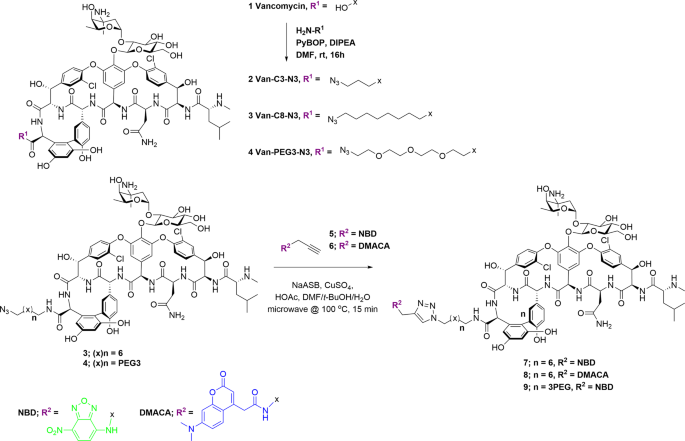
We have previously reported on the utility of azide-functionalised antibiotics and the Cu-catalysed azide−alkyne cycloaddition (CuAAC) reaction for linking antibiotics to fluorophores41,42,43,44,45. The CuAAC reaction is compatible with the multiple unprotected functional groups presented in antibiotics, in this case the many amine, hydroxyl, and amide groups on vancomycin. Other groups have reported successful CuAAC reactions with glycopeptides, including vancomycin, for the synthesis of modified glycopeptides46,47,48,49,50,51,52, glycopeptide dimers53,54, and glycopeptide fluorescent probes31. Therefore, we applied this strategy to the preparation of vancomycin analogues by synthesising three azide-derivatised vancomycins 2–4 containing different linkers (Fig. 1). We have previously reported on the preparation of vancomycin-nanoparticle conjugates by reacting the azide-functionalised vancomycin with alkyne-functionalised nanoparticles: these demonstrated enhanced binding affinity to bacteria and caused permeabilisation of the bacterial cell wall55. We are also investigating vancomycin-functionalised magnetic nanoparticles as capture probes for the development of a rapid bacterial diagnostic for blood- and other biological fluid-based infections56.

The C-terminus of vancomycin is amidated with amino-azido linker groups using standard peptide coupling reagents. The resulting azides 3 and 4 are then coupled with alkyne-derivatised fluorophores, NBD (green) or DMACA (blue), using the Cu-catalysed azide−alkyne cycloaddition reaction.
Antimicrobial activity of azido-vancomycin 2–4
As expected, the azide-vancomycin derivatives 2–4 retained antimicrobial activity against a representative section of Gram-positive bacterial strains including ATCC reference strains and clinical isolates of S. aureus, Streptococcus pneumoniae, Enterococcus faecalis, and Enterococcus faecium, with slight improvements in potency in some cases (Table 1). Azide-vancomycin 3 containing the octyl (8-C) linker was the most active, with MICs against most strains consistently two- to four-fold more potent than vancomycin (e.g. 0.5 µg/mL against MRSA, 32 µg/mL against VRE, 2 µg/mL against GISA compared to 1–2 µg/mL, ≥32 µg/mL, and 8 µg/mL respectively for vancomycin; our previous research into vancomycin analogues has shown lipophilicity at this position improves potency39). Both azido-vancomycin 2, with a shorter propyl (3-C) linker, and azido-vancomycin 4, with a more hydrophilic but longer polyethyleneglycol (PEG3) chain, showed comparable activity to vancomycin.
Conjugation with fluorophores
Fluorescent vancomycin probes were readily prepared by a single step synthesis with alkyne-derivatised fluorophores, using azido-vancomycins 3 and 4 as representative azides with different linker properties (both length, and hydrophobicity). To avoid undesirable steric or electronic interaction of the fluorophores with charged cell walls and minimise disturbance to antimicrobial activity, the fluorophores 7-nitrobenzofurazan (NBD) (green) and 7-(dimethylamino)-coumarin-4-acetic acid (DMACA) (blue) were selected, due to their comparatively low molecular weight and minimal (or positive) electronic charges (positive charge less likely to repel negatively charged bacterial surface). We have previously reported on the functionalisation of other antibiotic classes using alkyne-functionalised NBD 5 and DMACA 641,42,43,44,45.
Initially, CuAAC reactions of azido-vancomycin with the alkyne fluorophores gave poor yields of products 7–9 (Fig. 1). In previous literature reports, Nigam50 synthesised CRAMP-vancomycin via a CuAAC reaction (CuSO4·5H2O and sodium ascorbate (NaASB)) under microwave irradiation. CuAAC reaction under microwave irradiation has also been reported for the synthesis of a vancomycin hybrid49. However, this approach was not successful for our constructs, even when tris[(1-benzyl-1H-1,2,3-triazol-4-yl) methyl]amine (TBTA) ligand was also added: the reaction was sluggish and isolation of the product was not possible. Alternative copper sources such as Cu(OAc)2, CuI, and Cu(CAN)4PF6 were also evaluated, but with little success. Finally, reaction with CuSO4·5H2O (up to 0.5 equivalents) and sodium ascorbate at 40 °C in DMF/H2O (1:9) for 20 h gave good yields of the conjugation products50. Vancomycin is known to chelate copper, which may partially explain the poor yields of some of these reactions57.
The reaction was further optimised by using a solvent mixture of DMF/t-BuOH/H2O and adding acetic acid to accelerate the reaction, based on reports that acetic acid as a proton source accelerates the conversion of the C-Cu bond-containing intermediates (Cu(I) acetylide and 5-cuprated 1,2,3-triazole) into product, leading to reduction of by-products58,59. Given that electronic effects of the alkyne can influence the formation of the Cu(I) acetylide, optimisation of the quantities of CuSO4 and NaASB was evaluated separately for each fluorophore alkyne (5 required 0.2 eq. CuSO4 and 0.4 eq. NaASB, 6 required 0.5 eq. CuSO4 and 1.0 eq. NaASB). Adding excess catalyst did not improve the outcome of the reaction but increased unidentified by-products. Under the optimised microwave conditions, the CuAAC reactions in DMF/t-BuOH/H2O with CuSO4, NaASB, and HOAc as catalyst were completed within 15 min at 100 °C, providing the fluorescent vancomycins 7–9 in good yield. The excitation/emission wavelengths of Van-8C-Tz-NBD 7 (475/535 nm), when compared to Van-8C-Tz-DMACA 8 (375/480 nm) (Fig. S1), were generally more suitable for fluorescent microscopy as suitable filters are more readily available. Thus, azido-vancomycin 4 was conjugated with the NBD fluorophore 5 to generate another green probe 9.
Biological activity and quantification of bacterial staining with fluorescent vancomycin probes
Both fluorescent probes 7 and 8, with a C8 alkyl linker, showed improved (approximately two- to four-fold) antibacterial activity against multiple strains, when compared to the parent vancomycin (Table 1), again likely due to lipophilicity. Probe 9, with the PEG3 linker, retained the most similar antimicrobial activity to the parent vancomycin. We also compared the antimicrobial activity of probes 7, 8 and 9 to the commercially available vancomycin-derived fluorescent probes, Van-FITC (SBR00028, Sigma-Aldrich), and Van-BODIPY (V34850, Invitrogen™). Compared to Van-BODIPY, Probe 7 retained two-fold improved activity against all tested S. aureus strains, whereas Van-FITC did not possess antimicrobial activity at the highest concentration tested (32 µg/mL) (Table S3). The chemical stability of Probe 9 was assessed by analytical testing (HPLC) of old stock solutions that were dissolved in ddH2O and stored at −20 °C for over 3 years. No obvious degradation was evident, with purity >95% as determined by LC-MS using the same method employed to assess purity after synthesis. Furthermore, a one-year-old stock of Probe 9 showed identical antimicrobial activity as the fresh stock, indicating that the probe can be used for at least one year once dissolved (Table S3). For testing of Van-FITC and Van-BODIPY, they were both dissolved in DMSO according to their product manuals, instead of the water used for probes 7–9. Given that water is less disruptive to bacterial survival than DMSO, the ability to dissolve the new probes in water should cause less experimental interference.
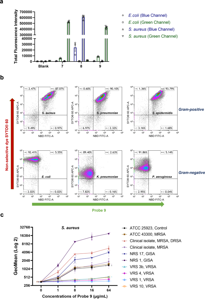
We next assessed the selectivity of the probes at distinguishing Gram-positive from Gram-negative bacteria. Traditionally, this is accomplished by Gram-staining, which relies on uptake and retention of a crystal violet stain to label the thick peptidoglycan layer of Gram-positive bacteria. However, many factors including the intactness and size of bacterial cells will affect the staining accuracy of this commonly used technique and it is not suitable for investigating viable bacterial cells because of a fixation step. Gram-negative-specific fluorescent probes such as polymyxin B-Cy360, tetramethylrhodamine-labelled tridecaptin A161, and Gram-positive-specific fluorescent probes such as vancomycin-Cy562, provide a new approach for labelling live Gram-negative bacteria and Gram-positive bacteria, respectively. In this study, the selectivity of vancomycin probes 7–9 were assessed. Flow cytometry analysis of bacteria labelled with the vancomycin fluorescent probes 7–9 clearly show selective labelling of S. aureus but not E. coli (Fig. 2a). The Van-DMACA probe 8 was somewhat less selective than the NBD-based probes (selectivity ratio for S. aureus over E. coli based on fluorescence uptake: 7 = 101:1, 8 = 25:1, 9 = 52:1). To further assess the specific labelling by probe 9, we analysed 3 Gram-positive and 3 Gram-negative bacteria co-stained with the green probe 9 and a non-specific cell permeable red nucleic acid dye SYTO® 60. SYTO® 60 stained all 6 strains while a shift in the FITC channel only appeared for Gram-positive strains, confirming selectivity between Gram-positive and Gram-negative bacteria (Fig. 2b).

a Labelling of S. aureus ATCC 25923 and E. coli ATCC 25922 with vancomycin probes 7–9 measured using flow cytometric analysis. Bacteria were treated with vancomycin probes at a concentration of 16 µg/mL (37 °C, 30 min), with fluorescence intensity of individual bacteria measured using flow cytometry. Positive events were gated on the histogram to estimate the mean fluorescence intensity (MFI). Total fluorescence intensity (TFI) of the gate was obtained by multiplying the MFI with the number of events on the gate. The data are presented as the mean ± SEM (n ≥ 3). b Labelling of bacteria with the Gram-positive selective vancomycin probe 9 and the non-selective cell permeable dye SYTO® 60 in various species using flow cytometry. Bacteria were treated with vancomycin probes at a concentration of 32 µg/mL (37 °C, 30 min) and SYTO® 60 (5 µM, 10 min on ice), then fluorescence intensity was measured using the flow cytometer. c Labelling of S. aureus with vancomycin probe 9 measured using flow cytometric analysis. Bacteria were treated with vancomycin probe 9 at different concentrations (37 °C, 30 min), then fluorescence intensity was measured using the flow cytometer. The data are presented as the mean ± SEM (n = 3).
We also compared fluorescent uptake of probe 9 by different S. aureus strains with varying degrees of vancomycin resistance, with vancomycin-sensitive and GISA strains showing a clear concentration-dependence (Fig. 2c). GISA and daptomycin-resistant strains, resistance phenotypes generally accompanied by thickened cell walls, showed moderately to highly increased levels of fluorescence (e.g. ~4- to ~18-fold increased fluorescence at 64 µg/mL of probe 9 for these strains compared to ATCC 25923). In contrast, VRSA strains showed substantially reduced staining (~6- to ~50-fold decreased fluorescence at 64 µg/mL of probe 9 for these strains compared to ATCC 25923), consistent with their different mechanism of resistance that reduces vancomycin's affinity (Fig. 2c and Table S1).
We also compared the staining ability of probe 7 and probe 9 to the commercial fluorescent probes using flow cytometry. The Van-BODIPY derivative performed best in staining S. aureus with the greatest intensity under the tested conditions. Probes 7 and 9 (as well as the one-year-old stock of Probe 9) were less effective, while the staining ability of Van-FITC was the worst (Fig. S2a). On the other hand, when incubated with the E. coli cells, Van-BODIPY stained a larger population of cells than the other probes, especially at 2 µg/mL, indicating a degree of non-specificity. In contrast, Probes 9 and Van-FITC did not show much staining with E. coli at either concentration, while Probe 7 was similar to Probe 9 at 2 µg/mL but had the highest E. coli labelling at 16 µg/mL (Fig. S2b).
Super-resolution confocal microscopy of bacterial staining with fluorescent vancomycin probes
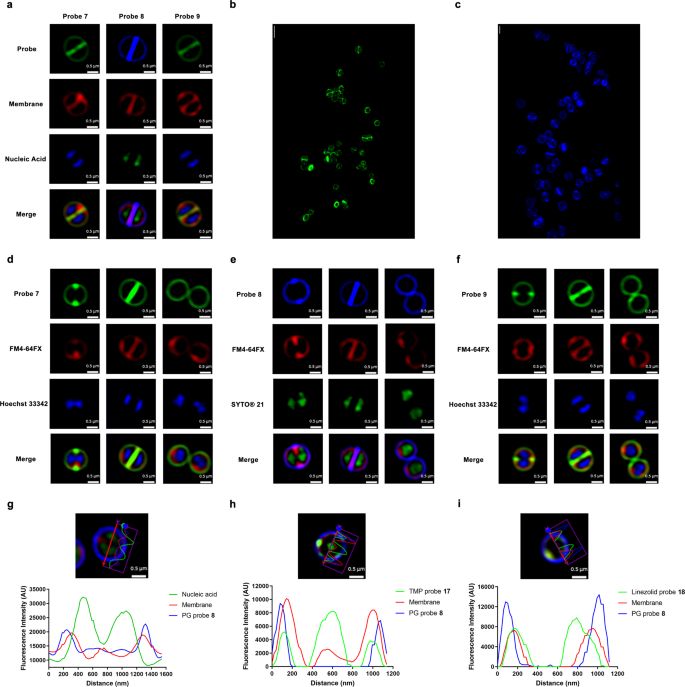
Super-resolution structured illumination microscopy (SR-SIM) is an essential tool to visualise bacterial cell structures, given their small size (approximately 1 µm) relative to human cells. Thus, S. aureus labelled with vancomycin probes 7–9 was fluorescently imaged using SR-SIM (Fig. 3a). Intense fluorescence at the dividing septum compared to the lateral wall confirmed binding to the nascent peptidoglycan Lipid II. The PEG linker (9) and the alkyl linker (7) did not lead to obvious differences in visualisation, although probe 7 was approximately 4-fold more active than probe 9 in MIC assays against the Gram-positive panel. The dividing septum ring was even more clearly visible when viewed using 3D-SIM (Figs. 3b, c and S3 and Supplementary Video). Similar labelling was seen in a range of other Gram-positive species and strains, including MSSA ATCC 25923, MRSA ATCC 43300, VISA NRS 1, VRSA VRS 4, Staphylococcus epidermidis ATCC 12228, Streptococcus pneumoniae ATCC 33400, Enterococcus faecium ATCC 35667 and Bacillus subtilis ATCC 6633 (Fig. S4). Notably, despite the relatively low levels of fluorescent uptake indicated by flow cytometry for VRSA, clear septal and cell wall labelling was still visible by microscopy.

a SR-SIM fluorescence imaging of S. aureus ATCC 25923 stained with probes 7–9. Bacteria were stained with Hoechst 33342 (blue, nucleic acid) or SYTO® 21 (green, nucleic acid) at 2.5 µM for 5 min on ice, followed by staining of FM4-64FX (red, bacterial membrane) at 5 µg/mL for 2 min on ice. Finally, vancomycin probes (16 µg/mL) were then added to the bacteria, left for 30 min on ice. Scale bar = 0.5 µm. b 3D-SIM of probe 7 (green) and c 3D-SIM of probe 8 (blue). Scale bar = 2 µm. d–f SR-SIM fluorescence imaging of S. aureus ATCC 25923 cell division with different probes. d Green; probe 7, Red; FM4-64FX (bacterial membrane), Blue; Hoechst 33342 (nucleic acid). e Blue; probe 8, Red; FM4-64FX (bacterial membrane), Green; SYTO® 21 (nucleic acid). f Green; probe 9, Red; FM4-64FX (bacterial membrane), Blue; Hoechst 33342 (nucleic acid). Scale bar = 0.5 µm. g–i Co-labelling of peptidoglycan (PG) labelling vancomycin probe 8 with trimethoprim and linezolid probes. The microscopy image shows SR-SIM fluorescence imaging of S. aureus ATCC 25923 with a cross-section measurement of fluorescent intensity in the accompanying graph. g Bacteria stained with probes 8 (16 µg/mL, blue, PG), FM4-64FX (5 µg/mL, red, bacterial membrane), SYTOX® green (2 µM, green, nucleic acid). h Combination of vancomycin-DMACA 8 (16 µg/mL, blue, PG) with TMP-4C-Tz-NBD probe 17 (64 µg/mL, Green), co-stained with FM4-64FX (5 µg/mL, red, bacterial membrane). i Combination of vancomycin-DMACA 8 (16 µg/mL, blue, PG) with linezolid-3C-Tz-NBD probe 18 (64 µg/mL, Green), co-stained with FM4-64FX (5 µg/mL, red, bacterial membrane). Scale bar = 0.5 µm.
Vancomycin probes have been used to study mechanisms of bacterial cell division via experiments including co-labelling with PG synthesis proteins63, fluorescence microscopy of vancomycin-labelled bacteria to show the dynamics of peptidoglycan assembly in ovococci17, and investigations of PG biosynthesis in B. subtilis25. Most bacterial cells grow and divided by building a new cell wall disc called the septum, which is the structure that forms in the middle of the mother cell by invagination of the cell membrane and ingrowth of the cell wall, leading to splitting of the mother cell into two identical daughter cells64. SR-SIM fluorescence microscopy successfully showed these stages of cell division, including ingrowth of the new PG, invagination of the cell membrane, and chromosome segregation, when labelled with the vancomycin probes, a membrane-labelling probe, and a nucleic acid-labelling probe, respectively (Fig. 3d–f).
Airyscan confocal super-resolution imaging was used to compare vancomycin probes 7 and 9, as well as the commercial probes Van-FITC and Van-BODIPY, for staining of S. aureus ATCC 25923 (Fig. S2c). With the exception of Van-FITC, all fluorescent probes showed good peptidoglycan binding ability. However, while Probes 7 and 9 ((as well as one-year-old stock of Probe 9) showed very strong fluorescence at the cell division septum (the site of Lipid II precursor incorporation into peptidoglycan) compared to the lateral wall, the Van-BODIPY probe appeared to label the cells more uniformly, indicating reduced selectivity of binding to nascent peptidoglycan. More intracellular labelling was also observed with the Van-BODIPY probe, further supporting that Probes 7 and 9 were more specific at labelling nascent peptidoglycan than Van-BODIPY (Fig. S2c). Van-FITC did not show good staining ability, consistent with its poor MIC and flow cytometry staining, with no fluorescence signal observed at 2 µg/mL and less clear labelling than other tested probes (Fig. S2c).
The vancomycin probes are able to spatially discriminate the peptidoglycan layer from the bacterial cell membrane as shown by cross-section measurements (Fig. 3g), which clearly show the peak of probe intensity outside of the peak intensity of a membrane-selective dye. Thus, vancomycin probes are potentially useful for helping to assess the target sites of other antibiotic probes such as those derived from linezolid and trimethoprim (TMP) with intracellular targets, by providing a reference point for the PG layer. Co-staining of S. aureus with the Van-DMACA probe 8 (blue) and trimethoprim-4C-Tz-NBD 1742 (green) or linezolid-3C-Tz-NBD 1841 (green) showed that the TMP and linezolid probes penetrated within the PG and bacterial cell membranes into the cytosol. Both TMP and linezolid probes generally showed co-localisation with the FM4-64FX (membrane dye) because the membrane dye was quickly endocytosed in S. aureus (Fig. 3h, i). These studies highlight the advantages of having additional colour versions of the antibiotic-fluorophore probes available for use in combination labelling studies.
Determining membrane permeabilisation in Gram-negative bacteria with fluorescent vancomycin probes: microscopy
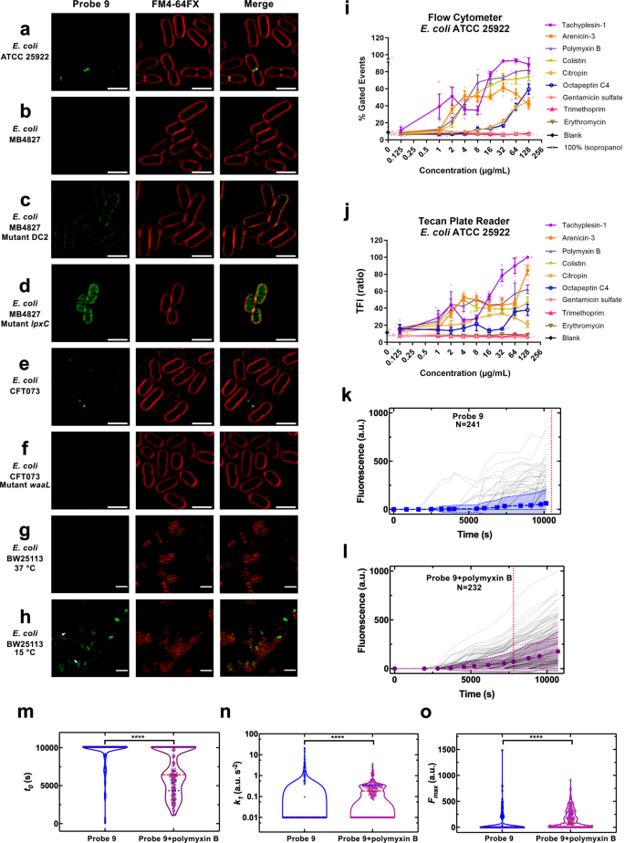
The OM of Gram-negative bacteria is an asymmetric bilayer that contains both phospholipid (inner leaflet) and LPS (outer leaflet). The LPS consists of Lipid A (a glucosamine-based phospholipid), a relatively short core oligosaccharide, and a distal polysaccharide (O-antigen)65. Due to the OM preventing access, vancomycin is inactive against Gram-negative bacteria (MIC > 64 µg/mL) (Table 2). Microscopy of E. coli (ATCC 25922, K12 (MB4827)) co-labelled with membrane dye FM4-64FX and vancomycin NBD probe 9 generally showed little evidence of vancomycin binding, as expected (Fig. 4a, b). However, occasionally cells exhibited strong fluorescence at the dividing septum just before cell division, where presumably the OM becomes 'leaky' as the cell begins to separate. The probe did not show any obvious labelling at the lateral wall. This inspired us to investigate the ability of the vancomycin probes to stain mutant bacteria with impaired OM.

SR-SIM fluorescence imaging of vancomycin probe 9 in E. coli strains ATCC 25922 (a), K12 (MB4827) (b), and CFT073 (e), and mutant E. coli (DC2 (c), lpxC (d) and waaL (f)). Bacteria were stained with probe 9 (32 µg/mL, Green) and FM4-64FX (5 µg/mL, red, bacterial membrane). Scale bar = 2 µm. g, h Visualisation of membrane permeabilisation at different temperatures. Airyscan confocal super-resolution imaging of probe 9 in E. coli strain BW25113. Bacteria were stained with probe 9 (32 µg/mL, Green) and FM4-64FX (5 µg/mL, red, bacterial membrane) at 15 °C or 37 °C. Arrows indicate cells labelled with probe 9 at dividing septum and peptidoglycan at 15 °C. Scale bar = 5 µm. i, j Determination of membrane permeabilisation in E. coli (ATCC 25922) using vancomycin probe 9. Bacteria were treated with compounds for 1 h at 37 °C, followed by labelling with the probe Vanco-NBD 9 (32 µg/mL), then left for 30 min at 37 °C. The samples were then measured by flow cytometer or Tecan plate reader. i Flow cytometry method. j Tecan plate reader method. The data are presented as the mean ± SEM (n ≥ 2). k–o Microfluidic single-cell analysis of probe accumulation in E. coli. Accumulation of vancomycin probe 9 in E. coli in M9 medium drug milieu at an extracellular concentration of 46 µg/mL in k the absence and l the presence of polymyxin B at an extracellular concentration of 1 µg/mL delivered to n = 241 and n = 232 individual E. coli, respectively. In both figures, data were collated from biological triplicate and fluorescence values were background subtracted and normalised by cell size. The symbols and shaded areas represent the mean and standard deviation of the corresponding single-cell values. The vertical dotted lines represent the time points at which the median of each dataset became larger than zero. The median remained zero throughout the entire experiments carried out with vancomycin-NBD 9 alone, hence the dotted line has been arbitrarily set at 11,100 s in k for comparison purposes only. Distributions of single-cell values for the kinetic parameters (m) t0, (n) k1 and (o) Fmax describing the accumulation of 9. The red dashed and blue dotted lines within each violin plot represent the median and quartiles of each data set, respectively. ****p value <0.0001. The fitting algorithm returned convergent transitions for the accumulation data of n = 46 (out of the 241 datasets available) bacteria treated with vancomycin-NBD 9 and for n = 161 (out of the 232 datasets available) bacteria cotreated with vancomycin-NBD 9 and polymyxin B. t0, k1 and Fmax values for single-cell accumulation profiles for which the fitting algorithm returned divergent transitions were arbitrarily set to 10,100 s, 0.01 a.u. s-2 and 0 for comparison purposes only. These values represent the time at which the experiments with both vancomycin-NBD and the combination of probe 9 and polymyxin B were terminated and the minimum values of k1 and Fmax measured across the two datasets, respectively.
Firstly, vancomycin probe 9 was incubated with permeable membrane mutants of E. coli, including a UDP-3-O-((R)-3-hydroxymyristoyl)-N-acetyl glucosamine deacetylase (LpxC) mutant MB4902 (deficient in LpxC therefore not efficient Lipid A production) and DC2 mutant CGSC7139 (deficient in osmoregulation of periplasmic oligosaccharide synthesis), comparing probe uptake to that of the parent strain, K12 (MB4827). Probe 9 was able to strongly label the mutant DC2 E. coli, indicating general penetration to the PG layer (Fig. 4c). The lpxC mutant E. coli also allowed probe 9 to label the PG, with apparently greater penetration to the cytoplasm compared to both control and the mutant DC2 strains (Fig. 4c, d). In MIC assays, both vancomycin and the probe 9 did show weak antimicrobial activity against the lpxC mutant E. coli (MIC = 64 µg/mL) (Table 2), confirming that LpxC deletion does allow for vancomycin penetration to the PG layer66,67,68.
Secondly, because mutation of O-antigen is known to alter antibiotic sensitivities65,69, an O-antigen mutant waaL E. coli with short length LPS (rough LPS mutant), was also stained with probe 9 and compared to the corresponding parent E. coli strain (CFT073) containing an intact O-antigen (Fig. 4e, f). In contrast to DC2 and lpxC mutants, probe 9 showed a similar lack of labelling in control and mutant O-antigen strains, indicating that O-antigen did not affect the permeability of the probe 9 to the PG under the tested condition (Fig. 4a–f).
Recent studies have suggested that vancomycin is able to inhibit growth of E.coli at low temperatures and that cold stress makes E. coli susceptible to glycopeptide antibiotics by altering outer membrane integrity70,71. To identify if the vancomycin fluorescent probe could detect this membrane damage caused by cold stress, we labelled E. coli cells grown at both 37 °C and 15 °C with vancomycin probe 9. Cells at 37 °C showed minimal labelling, which is consistent with our previous results. In contrast, cells grown at 15 °C showed substantial labelling at both the dividing septum and peptidoglycan (Fig. 4g, h), confirming changes of outer membrane permeability under cold stress.
Determining membrane permeabilisation in Gram-negative bacteria with fluorescent vancomycin probes: method development
The ability to visualise OM leakiness with the vancomycin probes triggered an investigation into their utility for quantifying OM damage caused by antibiotics. We developed two methods, using either flow cytometry or a plate reader for detection. E. coli ATCC 25922 was treated with 6 membrane-active antibiotics (tachyplesin-172, arenicin-373, polymyxin B74, colistin75, citropin76 and octapeptin C477) and 3 membrane-inactive antibiotics (gentamicin78: aminoglycoside 30S ribosome protein synthesis inhibitor; trimethoprim79: dihydrofolate reductase inhibitor, and erythromycin80: macrolide 50S ribosome protein synthesis inhibitor) at concentrations in range 0.125–128 µg/mL for 1 h, followed by labelling with the probe 9 (32 µg/mL). With the flow cytometry method (Fig. 4i), pure isopropanol was used as positive control. The fluorescence intensity of vancomycin probe-labelling following treatment with the membrane-active compounds increased with their concentration, while no damage was shown for the antibiotics acting by different mechanisms. For the membrane-active compounds, the level of OM disruption did not necessarily correlate with their MIC (Table S2), but structural classes appeared to exhibit similar trends. The β-hairpin antimicrobial peptides tachyplesin-181 and arenicin-373 led to increased fluorescence at the lowest concentrations, despite the cyclic lipopeptides polymyxin B and colistin possessing more potent MICs. The less potent cyclic lipopeptide octapeptin C477, and the ɑ-helical linear peptide citropin82 did require substantially higher concentrations before membrane damage was seen. These findings were further confirmed using a Tecan plate reader (Fig. 4j).
Single-cell microfluidics analysis of OM damage
Single-cell microfluidics provides an opportunity to bridge the qualitative analysis of tens of cells provided by confocal microscopy, and the bulk population analysis of millions of bacteria provided by flow cytometry and plate reader assays. Using our recently developed single-cell molecular accumulation platform hundreds of individual live bacteria, immobilised within channels in a microfluidic chip, can be monitored over time during the course of exposure to a fluorescent molecule45,83,84. Immobilised E. coli, exposed to the vancomycin probe 9, showed individual bacteria sporadically exhibited uptake of the probe, consistent with labelling during cell division (Fig. 4k). In contrast, when the OM permeabiliser polymyxin B was added, a much greater population of bacteria accumulated the probe (Fig. 4l). Next, we used our recently developed mathematical model to capture the phenomenology of drug accumulation in our experiment85 and inferred the time of onset of fluorescence (denoted t0), the rate of uptake (denoted by the rate constant k1), and steady-state saturation levels (denoted Fmax) describing the accumulation of the probe (Fig. 4m–o). We found that the vancomycin probe started to accumulate significantly faster in individual E. coli in the presence of polymyxin B (with a mean t0 of 6750 and 9374 s in the presence and absence of polymyxin B, respectively, ****). Moreover, the rate of uptake of the vancomycin probe was significantly greater in the presence of polymyxin B (with a mean k1 of 0.19 and 0.01 a.u./s2 in the presence and absence of polymyxin B, respectively, ****). Finally, also the steady-state saturation level of the vancomycin probe was significantly higher in the presence of polymyxin B (with a mean Fmax of 149 and 67 a.u. in the presence and absence of polymyxin B, respectively, ****). Taken together these modelling results affirm the experimental findings presented above.
Conclusions
Vancomycin fluorescent probes were designed and synthesised from azido-vancomycin building blocks using the CuAAC reaction. The fluorescent vancomycin probes retained good antimicrobial activity compared to previously reported vancomycin probes, possibly due to the use of small fluorophores with minimal electronic charges, and the procedure allowed for the facile introduction of fluorophores with different colours. We demonstrated the possibility of discriminating Gram-positive and Gram-negative bacteria using the fluorescent probe based on their selective labelling characteristics by flow cytometric analysis. Furthermore, when used for SR-SIM fluorescent imaging of Gram-positive bacteria, the vancomycin probes clearly showed the formation of freshly synthesised peptidoglycan and the probes were also able to spatially discriminate the peptidoglycan layer from the bacterial cell membrane. Finally, we demonstrated that the vancomycin probe 9 can be used to visualise membrane permeabilisation in Gram-negative bacteria caused by genetic mutations in the bacteria, by temperature stress, or by exposure to chemical compounds, with convenient flow cytometry or plate-based methods able to quantify the extent of outer membrane damage. Further studies should assess whether these selective staining characteristics are retained across a much larger range of Gram-positive and Gram-negative species and strains, while additional fluorophore colours would help expand the utility of the probes for co-labelling studies. These mechanism-specific methods (as the OM must become permeable to allow access to the peptidoglycan layer) could become a useful addition to the repertoire of tests commonly used to help define the mode of action of new antimicrobial compounds and the effects of bacterial pathway mutations.
Methods
General synthetic and analytical procedures
All materials, unless otherwise noted, were o...
Comments
Post a Comment